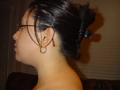
Monday, April 30, 2012

Day 30: A New Way of Thinking


This is the last day of blogging every day in April for the Cushing's Awareness Challenge.

For many months and like many Cushies, I have been feeling so STUCK and HOPELESS.  I felt like nothing I do seems to matter until I can get Cushing's off my back. There is just no need to keep rehashing what I already know: I am sick and uncured.  I KNOW!  Why must I remind myself of it every few minutes of every day?!


Someone recently reminded me that signs are all around us. Our minds must be open to see it, and our hearts must be open to take it in.  During this challenge, I have had many signs signaling to me what I need to do to move forward in my life.


I realize now that I have the world ahead of me, and it is my duty to pump as much life back into my daily living as I possibly can.

Time to learn to live with it because it is part of my life, at least for now.

From now on, I will work to think about myself in a much more positive light.

I will forgive myself more, and I will give myself a break.

I am doing the absolute best that I can each and every day. Some days I can do some things, and on the other days, I will rest and try again another day.

I must be more loving to myself.  I must drop all the guilt I feel about not being the kind of person/ friend/ mother/ wife I want to be, the kind of person that I used to be, not like this person I just met who is fabulous, or anything I wish I could do but can't quite do it now.

That is such a waste of effort!  I tires me, and I am already so exhausted. There is no reason to pile it on more.

I will make more effort to celebrate my caring spirit as I continue to help those around me who learn to live with Cushing's.

I don't do it enough, but I, too, must learn to deal with Cushing's with Moxie... every hour and every day it is with me.  It is the only way I know to continue moving forward and surviving this journey.

Onward,
Melissa

P. S. Just to compare, you can see day one's post for my Old way of thinking.  As for me, I'm not looking back.

Day 29: Dr Friedman's Everything Guide to Thyroid


The Everything Guide to Thyroid Disease: 

From potential causes to treatment options, all you need to know to manage your condition and improve your life

Purchase it on Amazon.com for only $11.

* ~ * ~ * ~ * ~ *

I am a patient of Dr. Theodore Friedman in Los Angeles, California, but like many, I traveled to see him when I lived in Texas.  Dr Friedman is a Cushing's expert and specializes in difficult-to-diagnose cases of the pituitary, adrenals and the entire endocrine system.  Dr Friedman sees five to ten patients with Cushing's each week and has for many years.
Doesn't make it sound that rare, now, does it?

If you are also interested in understanding the ins and outs of the thyroid glands -- overactive (hyperthyroidism), underactive (hypothyroidism), or self-attacking (Hashimoto's) -- I encourage to take a peak at this book.  It may be just what my doctor ordered.

If you would like more information about Dr. Friedman and his practice, read this excerpt from his website, GoodHormoneHealth.com:

So many of us believe that fatigue, weight gain, loss of libido and other problems are just symptoms we must learn to live with.  What if these symptoms are not the result of stress, diet, or aging, but are actually caused by a hormonal disorder?  Symptoms of hormone deficiency or excess may be subtle and difficult to diagnose.  Many hormonal problems are misdiagnosed as depression, especially in women.  


You know your own body better than anyone else, and you know when something is wrong.  Dr. Friedman is a compassionate, caring physician who will listen carefully to your concerns and work with you to establish a treatment plan.  As an experienced, board-certified endocrinologist and researcher, he has the capabilities to diagnose and treat even the most difficult hormonal problems.  Dr. Friedman has found that some of his patients suffer from undiagnosed pituitary or adrenal problems. These include many people suffering from Cushing's disease, which can present a baffling array of symptoms and is frequently misdiagnosed. Other patients may have pituitary or adrenal insufficiency, which has numerous symptoms and is equally hard to diagnose. Dr. Friedman is a world expert in these difficult-to-diagnose diseases and he welcomes inquiries from patients and their physicians.


Day 28: The Buffalo Hump


In the five years since I first learned of Cushing's, I have noticed many discussions among Cushies about their buffalo hump.  Folks just discovering Cushing's are particularly fixated with this discovery.

Who ever knew that an innocent deposit of fat could be a cause of such scorn, concern and distress?

I wrote about my self-guided path to diagnosis back in 2008. Today, I realized I haven't mentioned much about the buffalo hump since that post.  I now realize I have been remiss in not giving the buffalo hump the exposure it necessitates. The people want to know!

The buffalo hump -- listed among other many Cushing's symptoms -- was my a-ha symptom.

Excerpted from the post describing my self-guided path to diagnosis:

Not only did I experience nearly all of the symptoms on the list, I knew when I read the words "buffalo hump" that this was it. I just knew this is what I had.

Here is the buffalo hump picture I saw on the internet:


www.cushings-help.com


Here is my buffalo hump.
www.cushings-help.com
Copyright. Cushing's with Moxie. No reproduction without sourcing.


Copyright. Cushing's with Moxie.
No reproduction without sourcin

I searched the internet to see what else could cause a buffalo hump.
  • Cushing's (overproduction of cortisol caused by tumors in the pituitary or adrenal glands)
  • Long term use of steroids (when persons with normal cortisol production regularly take synthetic cortisol such as prednisone as medication to relieve the symptoms caused by another illness)
  • Pituitary tumor (could be source of Cushing's)
  • Hyperinsulinaemia (could be part of symptomology of Cushing's)
  • Morbid obesity (not on its own, usually part of the symptomology of Cushing's)
  • HIV and AIDS medications
  • Kyphosis (this is so easy to distinguish from a buffalo hump that I can't believe it made this list.  Kyphosis affects vertebrae between the shoulder blades and mid back, while a buffalo hump begins four inches below the hairline along the neck where the tops of the right shoulder and left shoulder meet in the middle.
    You can see kyphosis diagrams here and here.)
    NOTE: I measured my buffalo hump this way: hold up your right hand and tuck away your thumb. Place your four fingers sideways on your neck, with your pinkie touching the bottom of your hairline.  My index finger touches the top of my buffalo hump.
I looked up all the unfamiliar terms, and I knew they didn't apply. It could only be Cushing's.

My hump and me on a good day. We have a roller coaster relationship.Copyright. Cushing's with Moxie. No reproduction without sourcing.
So, to give my buffalo hump the credit it is due, I will share what I have learned about them over the years.

Q:  Is this a really a buffalo hump?
A:  If a person is asking about a buffalo hump, it is usually a buffalo hump.  Small or large, it is a hump. Accept this odd symptom especially when a patient has many other symptoms of Cushing's.

Q:  My doc says my hump is from obesity, but it looks like humps from diagnosed Cushies.
A:  Your doctor probably means well, but s/he hasn't seen as many Cushies as we have in the online community. In the past 60 months studying Cushing's and seeing my Cushie friends as real-time case studies, I have seen hundreds of patients think they have Cushing's, get knocked around in the diagnosis phase, only to have biochemically proven Cushing's with multiple tests (urine, saliva, blood) and/or ACTH-stained pathology from tumor(s) removed in pituitary surgery.
Also, see questions above.

Q:  My hump hurts. Does yours?
A:  Yes, mine kills me! My buffalo hump pulls at my neck making it hard to hold upright some days. I have spent hundreds of visits over the past 15 years at multiple chiropractors always looking for relief from the buffalo hump pain.  It never goes away, but sometimes I get relief for a few days.  Oh, how much money I must have spent in copays at the chiropractor!

I missed many days of work because this thing.  I would wake up and couldn't move my neck.  I couldn't lift my head off the pillow and would have to roll over onto my stomach to then push myself up off the bed just to get myself upright. It was not pretty.

Even today, I experience so much pain from this thing.  I feel a tension running from my neck down my right shoulder and no amount of heat/ ice combinations or stretching will ameliorate the pain. Typically, I have to take pain medication and lie down in order for relief to find me.

Also, my buffalo hump gets hard and soft!  It is so strange!  When the hump feels hard, it is usually causing me some pain.  When it is softer, sometimes I can press down and feel like I am not pressing on bone. It feels looser or softer.  I remember teling a Cushing's specialist about this, and he said, "I hear that all the time, but we don't know what causes the pain in a fatty hump or what makes it go up or down." Hmmphh. Neither do the patients!

Q:  Will it go away after pituitary surgery or when I am cured?
A:  Unknown.  Some say yes, some say no.  My presumption is to make sure your cortisol normalizes before really thinking you are gonna see any change.  That may take years after surgery (-ies).

Q:  My doc says it's a dowagers hump. My mom says it is poor posture. Could this thing really be a buffalo hump?
A:  They are wrong. A buffalo hump and kyphosis bump are located in different places on the spine. See discussion on kyphosis above the photo.  Also, when I take my side profile photos, I stand up super tall, shoulders back, head straight up as if a string is pulling my head up through the ceiling. Guess what? I still have a hump. It is not poor posture.

Q:  What do you do to hide your buffalo hump?
A.  To be honest, I don't hide my hump.  I can hardly drag myself out of bed or make it to the shower daily.  I have to focus on the big stuff, and for me, vanity flew out the window about 100 pounds ago.
However, I have seen others concerned about camouflaging their humps. Some wear their hair long to cover it, while others never wear a ponytail.  Some only wear collared clothes and never collarless clothes.  Again, for me, I can't be bothered by that thing!  Perhaps it helps that I can't see it every day, so really, I kinda forget about it.  Since you really can't do much about yours until your Cushing's is cured, I suggest you just forget it is back there, too. :)

~ * ~ * ~ * ~ * ~ * ~ * ~ * ~ * ~ * ~ * 


I saw this photo comparison for the first time this week. You can also see fat in the patient's neck and cheeks.

~ * ~ * ~ * ~ * ~ * ~ * ~ * ~ * ~ * ~ * 

Yes, this is a real place!  
Buffalo Hump Lake is located in south central Wyoming.

Day 27: Create Awareness by Telling Your Story


We build awareness about Cushing's when we share our own experiences. When others see themselves in our stories, we save that life and any others who learn from that person's experience.

Women & Diagnosis:
Help for Ladies Home Journal Article

Hello Moxie,

At the Pituitary Network Association, we are always hard at work trying to raise awareness for pituitary issues that affect patients like you, family members and physicians each and every day. And now, we need your help!

Most immediately, Ladies Home Journal Magazine is looking for women who were convinced something was medically wrong with them and who diagnosed their own illnesses despite baffled or even dismissive doctors. Ideally, this person should be diagnosed and currently being treated for their condition. If this situation sounds like you and you have a strong, working knowledge of your condition, and can speak about it in detail with the media, we would love to hear from you.

We are also looking to build our database of patient stories for future and ongoing media opportunities and requests. If you would be willing to speak openly about your story to the press, it could greatly help us raise awareness for others going through similar situations.

If you can help with either of the above requests, please e-mail us at info@pituitary.org. We will be in touch with further details.

Thank you!
The Pituitary Network Association is a strong advocate for those fighting pituitary tumors.

Source: Pituitary Network Association, www.pituitary.org

Day 26: Endocrine System Overview


In order to understand Cushing's, a patient must have a solid understanding of the endocrine system.  Today I share with you a very informative site describing the endocrine system.  This site has very cool diagrams; in fact, they are some of the best I've seen.  It includes information I have not seen on any other site that I have found.

The cellular stuff is hard for me to grasp, even after 60 months of living and studying this stuff.  However, I still try to learn.  Don't worry if you can't learn it all either.  For Cushies, learning new information and retaining what we learn or know is one of the most difficult mental tasks we face.  Always just do the best you can.


The Adrenal Glands: where they are and what hormones are made

How the Adrenal Glands Respond to Short-term and Long-term Stress


Sunday, April 29, 2012

Day 25: Another look at Cushing's

For those of you on Facebook, you may recall these Subject Slides going around.  The "What ___ say" part was always the same.  I saw some about Kansas, teachers, military intelligence, and everything else in between.  This is the one I created for Cushing's disease. I consider this one the "nice" one, as friends and family usually say things much more damaging, and doctors are in equal measures just as dismissive and heartless to the patient sitting in front of them.

Photo

Tuesday, April 24, 2012

Day 24: Pituitary Surgery and The Evolution from Open Transcranial to Fully Endoscopic Transnasal Surgery, and Beyond

I have several Cushie friends who recently had pituitary surgery with others headed there within the next few weeks.  For the Cushing's community, we celebrate these surgeries for the milestones that they are.  We rejoice!  After years of misdiagnosis and ill treatment from the medical community and even our loved ones, these are the days a sick person celebrates.

After having two unsuccessful pituitary surgeries myself, I just can't read enough about this process.  Please indulge me as I share yet another post about pituitary surgery.  If you, like me, want more, try this.

Today's Moment of Gratitude:  I'm glad I found out about my Cushing's after they stopped doing craniotomomies!


http://neurosurgery.stanford.edu/pituitary/review.html


Pituitary Surgery: The Evolution from Open Transcranial to Fully Endoscopic Transnasal Surgery, and Beyond
By Mohamed S. Kabil, M.D. and Hrayr K. Shahinian, M.D.


Historical background 

The development of pituitary surgery over the past century is largely credited to the pioneering work of Harvey Cushing in the early 1900s.1,2 Cushing accumulated extensive experience with various operative techniques including transcranial and transseptal-transsphenoidal approaches to the pituitary gland, but ultimately came to favor the transcranial procedure.3, 4 Schloffer in 1907 performed the first transsphenoidal pituitary tumor resection.5 For decades since the introduction of these techniques surgeons have debated which procedure provides the most complete resection of the pituitary lesion with the least risk of complications. Then in the 1960s Jules Hardy introduced intraoperative fluoroscopy and microscopy to transseptal-transsphenoidal pituitary surgery.1,6-8 The improved exposure afforded by these technologies allowed for complete removal of larger pituitary tumors, obviating the need for a complex transcranial operation in most patients. 

As a result, the transseptal-transsphenoidal approach came to be the procedure of choice for the surgical management of most pituitary lesions. Transcranial techniques were reserved for use in the resection of large tumors with extensive parasellar and suprasellar invasion. 

Recently, however, discussions regarding the most effective and least invasive way to perform pituitary surgery have been renewed. Developments in the field of endoscopic surgery have prompted surgeons to attempt endoscope-assisted surgery of the pituitary gland via the traditional transseptal approach.9-20 Recently, some surgeons have developed methods to perform transnasal-transsphenoidal pituitary surgery using only the endoscope for exposure.21-26 This procedure is proving to be equally if not more effective than the microscope as the primary imaging modality in pituitary surgery. 

For large pituitary tumors extending outside of the sella turcica, the transcranial approach offers the greatest chance of complete tumor removal. Often, a transcranial resection is reserved for the second stage of a two-stage operation.27,28 The first operation is performed via microscopic or endoscopic exposure of the pituitary tumor, during which time the bulk of the tumor is removed transseptally or transnasally. Any tumor extending outside the area visualized by the microscope/endoscope, or that is inaccessible during the first operation is then resected in a subsequent transcranial exploration. 

There are two major variations of the transcranial technique that are currently used for the resection of pituitary tumors: Midline Subfrontal Approach and the Oblique Subfrontal Approach. The operation is performed by first making an incision through the scalp, down to the skull on the side of greatest tumor extension. A craniotomy is then performed and the underlying dura incised; the frontal lobes of the brain are exposed and retracted to gain exposure to the tumor. Retraction must be kept to a minimum to avoid post-operative brain edema.27,29,30 Damage to the olfactory nerve can also occur during this operation, resulting in a decreased sense of smell.28,31,32 

Recently, minimally invasive techniques have also been extended to the transcranial approach. These are performed via an endoscopic transglabellar or supraorbital approach. The use of endoscopy allowed thorough visualization of all critical structures at the paramedian skull base without the need for a bicoronal scalp flap, bifrontal osteotomies, or brain retraction.33,34 

Microscopically assisted sublabial-transseptal-transsphenoidal pituitary surgery:

  • Indications: This procedure is indicated in the surgical management of pituitary tumors causing hypo- (not enough) or hyperfunctioning (too much) of the gland that have not adequately responded to medical treatment, or tumors growing into adjacent structures, evidenced by visual or other neurologic changes. Micro- and macroadenomas are amenable to resection by this method, including those with mild supra- and parasellar extension.1,35-39Large parasellar extensions of pituitary tumors, however, are more difficult to manage, as the lateral margins of the microscopic field of view are limited and blind removal of tumor extending beyond the sella is hazardous. Patients with extensive extrasellar extension of their pituitary tumors must be considered for two-stage procedures as described above.28,40,41,42
  • Surgical technique: While an assistant retracts the upper lip, a sublabial incision is made. Intranasal dissection is carried out separating the nasal tissue and removing a section of the central partition of the nose, forming a large "tunnel" through which the remainder of the procedure is conducted. An adjustable retractor is placed into this tunnel; its blades are advanced to the sphenoid sinus.

    The operating microscope is utilized for the remainder of the case, providing magnification and improved illumination of the limited operating field. The anterior wall of the sphenoid sinus is cautiously resected as it boarders the carotid arteries, cavernous sinuses, and optic chiasm. 43-45Once the anterior wall is removed, its mucosal lining is resected, exposing the posterior wall of the sinus (floor of the sella turcica). Benign adenomas usually do not invade the floor of the sella; however, expanding tumors may erode through the floor. If intact, the floor of the sella (posterior wall of the sinus) is removed. Dissection of this region determines the operative exposure, and is crucial for adequate visualization and thorough tumor removal. Deep to the floor of the sella is the dura. A sharp hook or blade is used to incise the dura, and through this incision specialized dissecting instruments are used to remove the tumor. Resection of tumor is carried out until all visually identifiable tumor is removed. As the sella is emptied of tumor, the boundaries of normal pituitary gland are identified.
The hole in the floor of the sella is then plugged using a fat graft from the abdomen, or muscle and fascia graft from the lateral thigh. This plug obliterates the space of the sphenoid sinus and helps prevent post-operative cerebrospinal fluid leakage.46 There are several models of graft design, in most cases a simple fat graft suffices.38,39,47-51 With the fat graft in place, the retractor is removed, the central bridge of the nose and flaps of tissue are repositioned, and the gum line incision is reapproximated using absorbable sutures. Both nostrils are packed with Vaseline-impregnated gauze strips. This packing remains in place for up to 48 hours postoperatively, absorbing any draining fluid and providing structural support to the nose as it heals internally. 

Fully endoscopic pituitary surgery 

With the advent of modern endoscopic equipment, momentum in the field of endoscopic pituitary surgery has stemmed from studies, which show endoscopes provide more comprehensive images of the pituitary gland and its surrounding structures than does the operating microscope.38,52 This in turn should allow for a more thorough tumor resection and fewer associated surgical complications. 

The clinical implications of these findings have been reflected in two separate studies of patients who underwent endoscope-assisted microscopic resections of pituitary tumors.15,47,52,53 These patients underwent a traditional microscopic transseptal-transsphenoidal approach to their pituitary gland tumor. Then, following what the surgeon believed to be complete tumor resection using the microscope, endoscopes were introduced into the pituitary region looking for residual tumor. In both series, an average of 40% of patients were found to have tumor left behind that was only discovered and resected during the endoscopic surveys. In other words, the microscope alone allowed for complete tumor removal in only 60% of patients. 

Improved tumor resection, elimination of intraoral and transseptal dissection along with reductions in operating time, recovery time and complications, have ushered in the completely endoscopic transnasal approach to the pituitary gland as the most recent phase in the evolution of pituitary surgery. 

  • Indications: Indications for fully endoscopic pituitary surgery are identical to those for the traditional transseptal-transsphenoidal microscopic approach.16,23-26
  • Surgical technique: The first step in the endoscopic procedure is to choose the appropriate endoscope. Preoperative physical examination of the nasal passages provides the surgeon with an idea of which endoscopes will be most appropriate. The surgeon must have scopes of varying diameters available, and must improvise intra-operatively depending upon the intranasal and skull base anatomy of the patient.

    Furthermore, every endoscope must be fitted with an irrigation sheath to clear the lens of blood or debris during dissection. This avoids the redundant removal and replacement of endoscopes for cleaning, which is both tedious to the surgeon and hazardous to the patient. The endoscope is attached to the grasping end of the holding arm, advanced into the right nostril, and used to conduct a brief survey of the anterior nasal passageway. The ultimate target of the endoscope is the sphenoid sinus. Therefore, the goal of the intranasal portion of the procedure is to create a passage to the sinus that is wide enough to accommodate the endoscope and accompanying instruments. This goal can be achieved rapidly, but should be meticulously and atraumatically performed, as bleeding from traumatized mucosa anteriorly can obscure visualization posteriorly.
The endoscope is advanced to the anteroinferior border of the middle turbinate. An elevator (dissecting instrument) is used to displace the nasal septum medially and middle turbinate laterally. A long straight suction device may also be introduced to clear the naris of any blood or mucoid secretions. As the nasal passage is widened, the holding arm is released and the endoscope advanced further posteriorly. Ultimately, the anterior wall of the sphenoid sinus is exposed, marking the extent of the intranasal dissection. The mucosal lining of the anterior wall of the sphenoid sinus is dissected away from the bone with a combination suction-cautery device and then lifted from the surface of the bone. Resection of the anterior wall of the sphenoid, the mucosal lining of the sinus, and the floor of the sella (back wall of the sinus) proceeds under endoscopic visualization. The surgical instruments are passed through the nostril, below the shaft of the endoscope, and into the surgical field to gain access to the sphenoid sinus and sella turcica. The same principles of awareness for the limits of dissection apply.16 Injuries to the cavernous sinuses, carotid arteries, optic nerves and chiasm are still possible if caution is not exercised while working within the sinus or sella. 

With the endoscope fixed in the sphenoid sinus, incision of the dura and removal of tumor proceeds as previously described. All imaging up until this point is provided by the 0º endoscope, which provides near complete visualization of the sella turcica, but only a limited view of the suprasellar structures. Therefore, once tumor resection is deemed complete, the 0o scope is replaced with a 30º endoscope. The 30º endoscope is advanced into the sella turcica and rotated clockwise and counterclockwise, thoroughly visualizing the supra- and parasellar regions. Such a comprehensive survey of these regions is not possible with the operating microscope.16,23-26 Tumor remnants in these areas are then removed, thereby eliminating sources of potential tumor recurrence. Resection is considered complete only after examination with the angled endoscope. A fat or muscle graft is then used to reconstruct the floor of the sella as previously described. No postoperative nasal packing is necessary. A small gauze sponge loosely taped beneath the nose collects any fluid that may drain from the nostril. Patients are discharged from the hospital within 24 to 36 hours of surgery. 

Endoscopic transcranial pituitary surgery: 

To minimize the deleterious effects of frontal lobe retraction and to avoid the use of facial incisions, approaches to these tumors have become progressively less invasive. Strategic placement of "keyholes" eliminates the need for excessive surgical manipulation without sacrificing exposure or outcome. 

The introduction of Endoscopic skull base surgery has allowed the resection of these tumors thru two minimally invasive approaches that involve placing a small (2cm) incision either within the skin crease in the bridge of the nose or within the hair of the eyebrow, depending on the exact location of the tumor.34 

  • Indications: For large pituitary tumors extending outside of the sella turcica, the transcranial approach offers the greatest chance of complete tumor removal. Often, a transcranial resection is reserved for the second stage of a two-stage operation.27,28
Endoscopic transglabellar approach to the pituitary gland 

The adaptation of rigid endoscopy to the transglabellar approach broadens the available surgical exposure without the introduction of additional dissection or retraction. Endoscopes of varying angles of view provide a panoramic perspective of the relevant surgical anatomy and allow for thorough evaluation of the extent of intracranial and extracranial disease. The maneuverability of the endoscope allows the surgeon to position it directly at the level of dissection, effectively reducing the viewing and operating distances. Endoscopic imaging thereby facilitates complete tumor resection via a minimally invasive technique. This technique obviates the need for a bicoronal scalp flap or an extensive bifrontal craniotomy without diminishing visualization of the paramedian skull base. 

  • Surgical technique: The patient is placed supine on the operating room table and the head of the bed is slightly raised. Following the induction of general anesthesia, the patient's neck is extended approximately 15° and the head is fixed in place using a three-pin clamp. Thus positioned, the frontal lobes will fall away from the floor of the anterior once cerebrospinal fluid (csf) is drained. The frontal and nasal areas are cleansed with an aqueous antiseptic solution and then draped.

    The base of a pneumatically powered endoscope holding arm is fastened to the operating room table opposite the surgeon; the arm extends over the patient. A 4.0 mm 0° rigid endoscope is attached to the holding arm.

    A 4 cm incision is made between the medial ends of the eyebrows, crossing the nasion in a skin crease. The skin flap is developed in a subcutaneous plane and retracted superiorly. The glabellar periosteum is elevated separately and retracted inferiorly for further use as a pedicled pericranial flap in reconstruction of the skull base. A small burr hole is placed in the frontal bone and the outer table of the frontal sinus is osteotomized. Once the sinus cavity is exposed, its mucosa is resected and the nasofrontal ducts are obliterated. A burr hole is then placed in the posterior wall of the sinus and a second bone flap is removed, revealing the underlying dura. The craniotomy can be extended laterally over the orbital roofs as dictated by the surgical anatomy of the tumor. An incision is made in the dura and csf is liberally drained. With relaxation of the frontal lobes, the endoscope is advanced intracranially along the floor of the anterior fossa between the olfactory tracts. Endoscopic survey reveals the degree of intracranial tumor spread. Extrasellar extensions of tumor are then exposed and removed from this superior approach.

    Prosthetic dural graft material can be used to ensure a watertight seal in dural repair. The pedicled periosteal flap is interposed between the dura and the paranasal sinuses to reconstruct the base of the skull when appropriate. The nasoglabellar bone flap is repositioned using absorbable microplates and screws. The skin incision is closed with careful attention to the aesthetic repair. The patient is monitored in the intensive care unit until neurologically stable and thereafter transferred to the ward until discharge from the hospital.33
Endoscopic supraorbital (eyebrow) approach to the pituitary gland 

Involves placing an incision within the hair of the eyebrow, performing a 1-1.5cm keyhole supraorbital opening and advancing the endoscope along the floor of the anterior cranial fossa underneath the frontal lobe. This approach allows a panoramic visualization of the ipsilateral anterior fossa and a partial visualization of the contralateral anterior fossa. 

  • Surgical technique: Subsequent to the skin and soft tissue incision, a 1.5cm craniectomy is performed. The dura is incised and cerebrospinal fluid drained. The endoscope is introduced thru the keyhole and advanced between the frontal lobe and the floor of the anterior skull base all the way to the tumor. A panoramic view of the tumor is displayed on a flat screen. Using a combination of a custom designed bipolar electrocoagulation system and a micro cavitron ultrasonic aspirator the tumor is gradually resected. This allows a complete and total resection of virtually most anterior skull base tumors through minimally invasive techniques with minimal or no brain retraction. More than 90% of all patients undergoing these procedures are discharged from the hospital within 48 hours.
Conclusion: 

The evolution of pituitary surgery over the past decade has been characterized by a progressive trend toward less invasive surgical approaches to the gland. Innovations in medical technology have, in part been responsible for these advances. The transcranial method was abandoned for the transseptal technique when surgeons documented their ability to achieve equal surgical results without the need for removal of the skull or retraction of the brain. The endoscopic transnasal approach offers even less invasive access to the pituitary gland and surrounding area, in addition to providing better intraoperative imaging of the region. Evidence suggests that complication rates and surgical outcomes of endoscopic pituitary surgery compare favorably to those that have been reported in large series of patients who have undergone microscopic transseptal pituitary surgery.18-21 More extensive data over longer periods of follow-up will further substantiate these trends. It is clear that transnasal endoscopic pituitary surgery represents significant progress in the surgical management of pituitary disease. Furthermore, the endoscopic transcranial approach whether transglabellar or supraorbital to the pituitary gland is an effective adjunct and a far less invasive alternative to the traditional transcranial approaches. 


Glossary of Terms
  • Transcranial: Surgical exposure for pituitary surgery by way of opening the skull (Craniotomy), and retracting the front of the brain to access the pituitary gland
  • Transseptal: Surgical resection of a portion of the central partition of the nose to access the sphenoid sinus. (exposed either transnasally or through sublabial incision)
  • Transglabellar: Surgical exposure for pituitary surgery by way of opening the skull through an opening made at the glabella (the forehead)
  • Supraorbital: Above the orbit (eye socket)
  • Sphenoid sinus: Sinus (cavity lined with mucosa) that lies directly behind the nose and in front of the pituitary gland: the back wall of which makes up the anterior wall of the sella tursica.
  • Transsphenoidal: Surgical dissection through the sphenoid sinus to access the pituitary gland.
  • Transnasal: Surgery through the nostril providing access to the sphenoid sinus and pituitary gland.
  • Sublabial: Surgical incision below the lip, above the front teeth in the gum line, providing access to the nostril, or followed by splitting the palate (bone) to eventually access the sphenoid sinus.
  • Sella Tursica: Bony structure at the base of the skull in which the pituitary gland rests.
  • Intrasellar: Within the sella tursica.
  • Subsellar: Extending below the sella tursica.
  • Suprasellar: Extending above the sella tursica.
  • Parasellar: Extending into the area surrounding the sella tursica: (beside, behind or in front of)
  • Adenoma - Benign tumor - referring to a non-functioning mass in the pituitary gland (this paper)
  • Microadenoma: Small adenoma: Less than 10mm in size.
  • Macroadenoma: Large adenoma: Greater than 10mm in size.
  • Anterior: Pertaining to being in front of another structure - toward the front of the body
  • Posterior: Pertaining to being behind another structure - toward the back of the body
  • Medial: Toward the midline of the body
  • Lateral: Toward the periphery of the body (away from the midline)
  • Carotid arteries: Arteries which carry blood up through the neck, eventually supplying the brain with its major blood supply: these vessels are also found within the cavernous sinus
  • Cavernous sinus: Area adjacent to the sphenoid sinus containing several vital nerves and blood vessels
  • Dura: Thin membrane surrounding the brain, which acts as a bag to contain the cerebrospinal fluid.
  • Cerebral Spinal Fluid/Leak: Clear fluid which surrounds the brain/a hole in the dura which allows the leakage of this fluid (the dura must be opened to access the pituitary gland)
  • Endoscopy/Endoscopic Surgery: Surgery performed using small illuminated lenses on long rods connected to cameras to magnify/better visualize an operating field.
  • Fluoroscopy: Video x-rays
  • Frontal Lobes/edema: The region of the brain in the front of the head which lies above and in front of the pituitary gland/a swelling of some tissue due to injury and accumulation of fluid
  • Indication: Scientific/medical reason for performing a certain procedure
  • Microscopy: Using an operating microscope to better visualize a small operating field or perform delicate surgery
  • Olfactory Nerves: Nerves which connect to the nostrils to provide one's sense of smell
  • Optic Chiasm: Area where optic nerves cross over, located just above the pituitary gland, which can be compressed by pituitary tumors
  • Optic Nerves: Nerves which connect to the eyes providing one's sense of sight
  • Resected: Cut out or removed surgically
  • Turbinate (superior, middle, inferior): Bony prominences in the nasal passage way
Click here for article references.

Day 23: File It Away...Solu-Cortef has a new National Drug Code

SCENARIO:  You are a Cushie headed for pituitary or adrenal surgery and you are taking care of business! You drop off your prescription for Solu-Cortef and hydrocortisone at the pharmacy. When you return in an hour, the staff informs you they can't find Solu-cortef in the system.  They no longer offer it, so the pharmacy can't fill it.

WHAT? You panic. You have to have this medication. A post op Cushie and her Solu-Cortef injectible is like a diabetic and his glucagon injection.  In a crisis, you could die without it.

What are you gonna do?  Call your doctor?  Doctors don't mess with this minutia. Nurses/staff don't know even know what Cushing's is, how it is treated. They do not know how to navigate the system at this level. After many exasperated phone calls and voice mails back and forth... nothing. No medicine for you.

What are you gonna do?  Put on your super hero cape.
It's up to you, the patient, again.
So what are you gonna do?

SOLUTION:  You fall back on the details you learned from a patient with Cushing's... me or any other who has shared any bit of information with you.  You have voraciously read this blog and many others over the years.  You read and you learn.  You file them away in that brain of yours, never quite sure you could recall anything up there when needed because the Cushing's dulls your brain so much. Either you remember, or a Cushie friend who read and remembers does.

PATIENTS KNOW BEST.

These are the kinds of details... the minutia... that Cushing's patients must wade through to stay alive. Make sure you know all that you can know about your health and disease. I promise that you will save your own life on many, many occasions.

~ * ~  * ~  * ~

Not sure why Solu-Cortef is important? You can read about Cushie troubles.  

~ * ~  * ~  * ~

 Solu-Cortef has a new National Drug Code (NDC)

We have recently been informed that Pfizer, the manufacturer of Solu-Cortef®, in the convenient "acto-vials," has removed the preservatives from their product, so have been issued a new NDC number by the FDA. The number is 0009001103.

This NDC number change has apparently made it difficult for pharmacies to find the product in their computer systems, resulting in erroneously informing people that it is no longer available for prescription.

The product is available, but with a new code. The number is 0009001103.

If your pharmacy is having trouble obtaining these products, please instruct them to call Pfizer's Customer Service at 1(800) 533-4535.

Other possible contact numbers for Pfizer are 1(800) 821-7000 or patient services at 1(888) 691-6813.  Toll-free: 866-227-3737.  Phone: 908-364-0272


Day 22: Rare Hormone Disorder Brings New Hampshire Woman To OSUMC


Day 21: Magic Foundation hosts Cushie Convention


A patient with Cushing's often travels a long and lonely road to diagnosis. Surrounded by friends and family who mean well but don't quite get it, a Cushie longs to connect with others who knows what it is like to fight while living with  Cushing's.

Well, here's your chance!  

Many Cushing's patients attend the annual Magic Foundation convention. While the Magic Foundation is known for advocating for children and adults with growth hormone deficiencies, they also have Cushing's patients at the MAGIC Foundation.  Stacey, the group's moderator, shares her personal story about Cushing's.

Anyhoo.

MAGIC offers an educational program for adults who are affected with Growth Hormone Deficiency and/or other endocrine disorders.
The  
MAGIC Foundation 
presents: 

The 7th Annual Convention  

for Adults with  

Pituitary Disorders  

Including:

Adult Growth Hormone Deficiency  
Panhypopituitarism 
and Cushings!

Thursday - Sunday 
July 19-22, 2012 
The Westin Lombard   
Yorktown Center 
Lombard, Illinois
Westin Hotel in Lombard
(subdivision area of Chicago)
Chicago, Illinois


Click through for convention program and online registration form.

~ * ~ * ~ * ~ * ~
  
Enjoy the fun. I won't be able to make it this year, but one day I promised myself, I will. It's too good of a party to pass up.